






The heart works continuously to move blood throughout the body, and when its function changes, physicians rely on detailed imaging to understand what is happening. An echocardiogram is a tool for examining the heart. It offers a clear view of the heart’s structure and movement without requiring surgery. For people living with heart failure or those recently diagnosed, this test can become a regular part of ongoing care.
What Is an Echocardiogram?
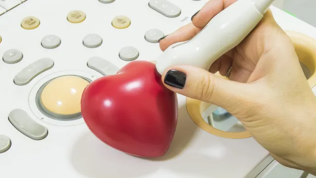
An echocardiogram is a painless test that uses sound waves to create moving pictures of the heart. These sound waves, known as ultrasound, bounce off the heart’s chambers and valves to produce images on a screen. A technician places a small device called a transducer on the chest, and this device sends and receives the sound waves. The resulting images show how the heart beats, how blood flows through it, and how well the valves open and close.
The test does not involve radiation, which sets it apart from some other imaging methods. Most echocardiograms take between 40 and 60 minutes to complete. During the procedure, you typically lie on an exam table while the technician moves the transducer to capture different views. The recorded images are then reviewed by a physician who studies the heart’s size, shape, and pumping strength. The test is noninvasive and repeatable; it serves as a practical way to track changes in heart function across months or years.
What Are the Types?
Several forms of echocardiography exist, and each one serves a specific purpose depending on what the physician needs to observe. The choice of test depends on the specific questions a physician wants to answer about your heart. There are many different methods for utilizing echocardiograms, including:
- Transthoracic Echocardiogram (TTE): This is the most common version. The transducer rests on the outside of the chest, making it a straightforward option for many patients.
- Transesophageal Echocardiogram (TEE): For this test, a thin tube with a transducer is guided down the esophagus. Because the esophagus sits close to the heart, this method produces highly detailed images, which can be useful when a standard chest scan does not provide enough clarity.
- Active Stress Echocardiogram: This version records images before and after physical activity, such as walking on a treadmill. It helps show how the heart responds when it works harder.
When exercise is not suitable, medication may be used to simulate the effects of activity for a stress test.
What Aid Does One Provide?
An echocardiogram can reveal a wide range of details about heart health. It may measure how much blood the heart pumps with each beat, and this measurement helps classify the type and severity of heart failure. The test can also show weakened or thickened heart muscle, as well as valves that do not function properly.
Physicians use echocardiogram results to track whether the heart is stable, improving, or declining over time. Medication or lifestyle changes may be introduced, and follow-up scans help show whether those adjustments are having the intended effect. The test can also support planning for procedures or devices when the situation calls for them. By providing a clear view of heart function, echocardiography helps care teams tailor treatment to each person’s changing needs.
Meet With a Cardiologist
A cardiologist reviews echocardiogram results alongside your symptoms, medical history, and other test findings to form an understanding of your heart health. This specialist is able to explain what the images mean, and they can outline the monitoring schedule that fits your condition. Regular follow-up allows small changes to be noticed early, which supports steady and thoughtful care.