





Skin cancer diagnosis describes the process dermatologists use to evaluate unusual spots, moles, or patches of skin. It follows a clear sequence, and each stage builds on the one before it. The process typically involves a visual examination of the skin, followed by a biopsy when a suspicious area requires further investigation. By identifying abnormal skin changes early, healthcare professionals can diagnose skin cancer more effectively and determine the most appropriate treatment. Here’s more information about how dermatologists diagnose this condition:
Gathering History and Visual Clues
A skin exam typically begins with a conversation. Your dermatologist asks about your personal and family history of skin cancer, and they may inquire about past sunburns. Additional risk factors include having light-colored skin and smoking cigarettes. If you noticed a recent change in a spot, mention it at this stage. The dermatologist also examines your skin from head to toe. They may check areas people sometimes overlook, such as the scalp and the soles of the feet. Many dermatologists use the ABCDE guide. The list below explains each letter:
- Asymmetry: One half is different from the other
- Border: The edges look ragged or blurred
- Color: Several shades appear within one spot
- Diameter: The spot measures more than approximately 6 millimeters
- Evolving: The spot changes size, shape, or color
Using Imaging and Magnification Tools
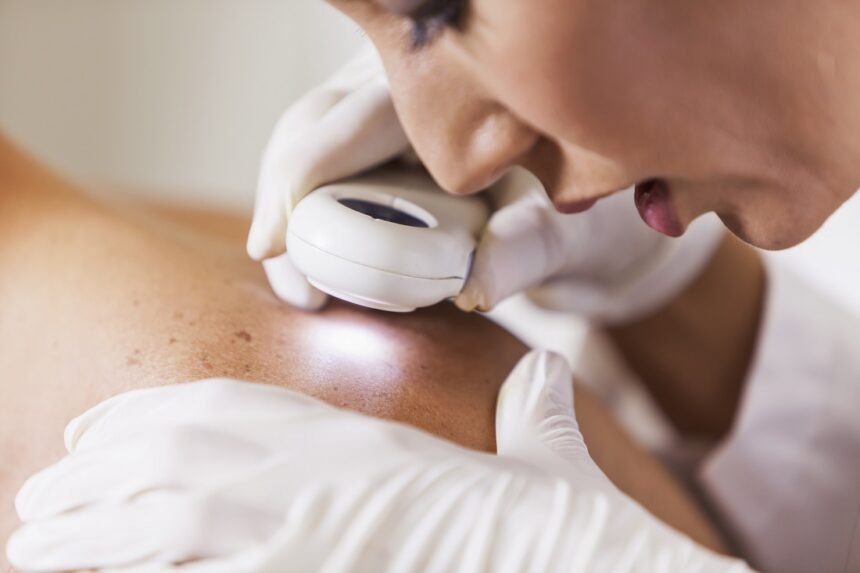
Dermoscopy plays a key role in many skin exams. A dermatoscope combines a magnifying lens with a built-in light, and it helps reveal additional details. The device may also connect to a camera. Clinics save the images, and the dermatologist reviews them at later visits. When a spot shifts in size or color, the saved photos make that change easier to see. Dermatologists may also use total body photography for patients with many moles. This method captures high-resolution images of large skin areas, and software compares new photos with earlier ones. The program flags new moles or subtle shifts that a quick glance might miss.
Confirming Results Through Biopsy
A biopsy provides the information a dermatologist needs to confirm a diagnosis. To perform the procedure, the dermatologist numbs the area with a local anesthetic. They remove a small tissue sample, and they send it to a laboratory for examination. Dermatologists may choose from several biopsy methods depending on the characteristics of the suspicious area. A shave biopsy uses a thin blade to remove the upper layers of skin, while a punch biopsy uses a circular tool to collect a deeper tissue sample. The dermatologist selects the most appropriate method based on the size, depth, and location of the suspicious spot.
At the laboratory, a specialist examines the tissue under a microscope. If the results confirm skin cancer, the doctor may recommend further testing to determine whether the cancer has spread. These tests sometimes include a lymph node biopsy or imaging scans; the healthcare team explains the purpose of each procedure.
Consult a Dermatologist
Dermatologists diagnose skin cancer through a series of connected steps. A consultation and full-body examination typically come first, while magnification tools provide additional detail. When a spot requires closer evaluation, a biopsy and laboratory analysis help confirm the diagnosis. Regular skin checks provide your dermatologist with a valuable baseline. Review when you last had a full-body examination, and note any spots that have changed. If you notice something new or unusual, schedule an appointment with a board-certified dermatologist.